vol. 107 no. 30 > Dharmendra S. Modha, 13485–13490, doi: 10.1073/pnas.1008054107
Network architecture of the long-distance pathways in the macaque brain
 Dharmendra S. Modha and Raghavendra Singh
PNAS July 2010
- Proceedings of the National Academy of Sciences of the United States of America,
vol. 107 no. 30 > Dharmendra S. Modha, 13485–13490, doi: 10.1073/pnas.1008054107
Network architecture of the long-distance pathways in the macaque brain
Dharmendra S. Modha and Raghavendra Singh
Start of Abstract
Understanding the network structure of white matter communication pathways is essential for unraveling the mysteries of the brain's function, organization, and evolution. To this end, we derive a unique network incorporating 410 anatomical tracing studies . . . network consists of 383 hierarchically organized regions spanning cortex, thalamus, and basal ganglia; models the presence of 6,602 directed long-distance connections; . .
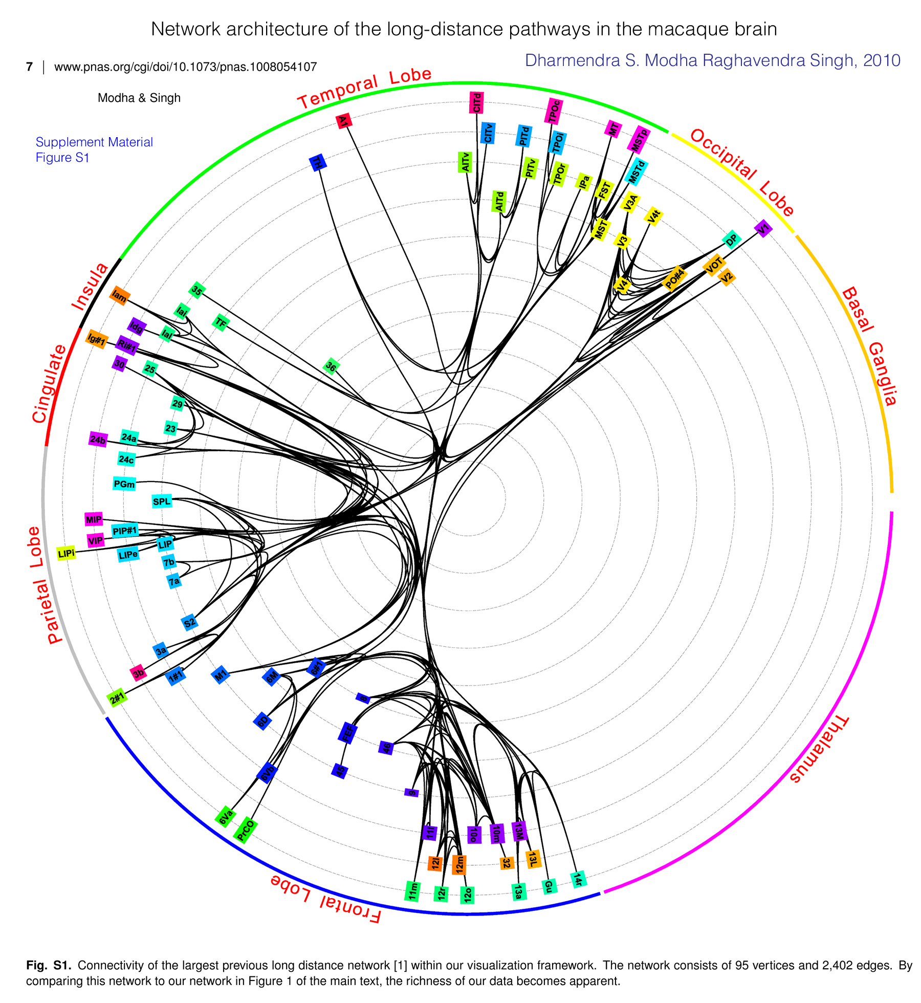
The Supplemental Material!! is especially rich in visualizations of various types. The images shown here have all of the hierarchies with a bit of a tight fit. There are many images showing the individual hierarchies.
The images showing the "bundling" of the fibers shows what the graph would look like with no bundling and with two intermediate steps. In the image to the left - Figure 1 from the paper - you can see all of the possible connections, but, the fibers that run parallel are "bundled" to reduce clutter.
One thing the supplementary material does not have is a vector version of Figure 1. Figure S1. is a vector version.
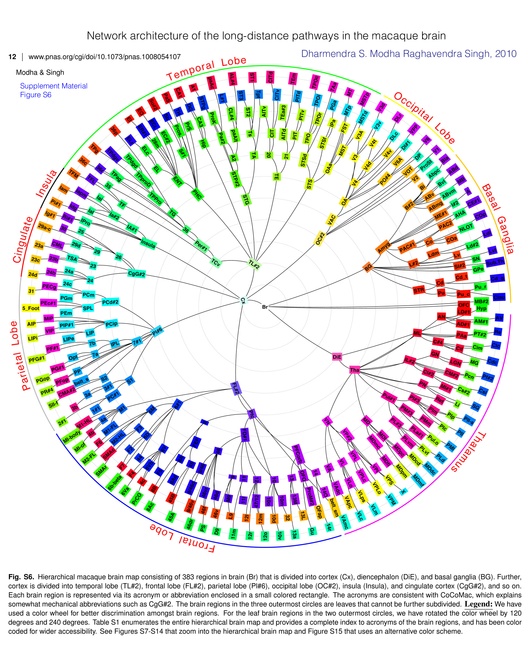
And the second image Figure S5. is is the whole hierarchy.
 Irimia: Similar Image for humansCircular representation of human cortical. . .Andrei Irimia, Micah C. Chambers, Carinna M. Torgerson, and John D. Van Horn |
The CoCoMac data base seems to be defunct, but, Kotter 2005 gives context. For example, GM-Definition is the General Map, as in the acronym map, general map and regional map. BTW, below is a similar image for humans by Andrei Irimia
Circular representation of human cortical. . .
| 1 | Br | According to GM-Definition |
| 2 | DiE | Diencephalon according to GM-Definition alamus |
| 3 | Hyp | Hypothalamus |
| 4 | Tha | Thalamus |
| 5 | AN | Anterior nuclei of the thalamus |
| 6 | LD#1 | Laterodorsal nucleus (thalamus) |
| 7 | AV | Nucleus anterior ventralis thalami |
| 8 | AM#1 | Nucleus anterior medialis thalami |
| 9 | AD#1 | Nucleus anterior dorsalis thalami |
| 10 | ML | Midline nuclei of the thalamus |
| 11 | Re | Nucleus reuniens thalami |
| 12 | PT#2 | Nucleus parataenialis thalami |
| 13 | PAa | Nucleus paraventricularis thalami, pars anterior |
| 14 | C#4 | Nucleus centralis thalami |
| 15 | Clc | Nucleus centralis latocellularis thalami |
| 16 | Cim | Nucleus centralis intermedialis thalami |
| 17 | Cif | Nucleus centralis inferior thalami |
| 18 | Cdc | Nucleus centralis densocellularis thalami |
| 19 | GN | Metathalamus (Geniculate Nucleii) |
| 20 | MG | Corpus geniculatum mediale |
| 21 | LGN | dorsal Lateral geniculate nucleus |
| 22 | IL#2 | Intralaminar nuclei of the thalamus |
| 23 | Pf#2 | Nucleus parafascicularis thalami |
| 24 | Pcn | Nucleus paracentralis thalami |
| 25 | CM#2 | nucleus centrum medianum (thalamus) |
| 26 | Cl#2 | Nucleus centralis lateralis thalami |
| 27 | Csl | Nucleus centralis superior lateralis thalami |
| 28 | Cs#2 | Nucleus centralis superior thalami |
| 29 | MI#1 | Massa intermedia |
| 30 | PN | Posterior Nucleii of Thalamus |
| 31 | SG | Nucleus suprageniculatus thalami |
| 32 | Li | Nucleus limitans thalami |
| 33 | Ret | Nucleus reticularis thalami |
| 34 | Pul#1 | Nucleus pulvinaris thalami |
| 35 | PI#3 | Nucleus pulvinaris inferior thalami |
| 36 | PIl-s | Nucleus pulvinaris inferior thalami, shell of the lateral subdivision |
| 37 | PIp | Nucleus pulvinaris inferior thalami, posterior subdivision |
| 38 | PIm | Nucleus pulvinaris inferior thalami, pars medialis |
| 39 | PIl | Nucleus pulvinaris inferior thalami, lateral subdivision |
| 40 | PIc | Nucleus pulvinaris inferior thalami, central subdivision |
| 41 | PM#3 | Nucleus pulvinaris medialis thalami |
| 42 | PMm | Nucleus pulvinaris medialis thalami, medial division |
| 43 | PMl | Nucleus pulvinaris medialis thalami, lateral division |
| 44 | Pul.o | Nucleus pulvinaris oralis thalami |
| 45 | PL#3 | Nucleus pulvinaris lateralis thalami |
| 46 | PLa#1 | Nucleus pulvinaris lateralis thalami pars alpha |
| 47 | PLd | Nucleus pulvinaris lateralis thalami, dorsal division |
| 48 | PLvl | Nucleus pulvinaris lateralis thalami pars ventrolateralis |
| 49 | PLvm | Nucleus pulvinaris lateralis thalami pars ventromedialis |
| 50 | MD | Nucleus medialis dorsalis thalami |
| 51 | MDdc | Nucleus medialis dorsalis thalami, pars densocellularis |
| 52 | MDcd | Nucleus medialis dorsalis thalami, pars caudodorsalis |
| 53 | MDl | Nucleus medialis dorsalis thalami, pars lateralis |
| 54 | MDpc | Nucleus medialis dorsalis thalami, pars parvocellularis |
| 55 | MDmf | Nucleus medialis dorsalis thalami, pars multiformis |
| 56 | MDmc | Nucleus medialis dorsalis thalami, pars magnocellularis |
| 57 | MDpm | Nucleus medialis dorsalis thalami, pars paramediana |
| 58 | MDfi | Nucleus medialis dorsalis thalami, pars fibrosa |
| 59 | VN | Ventolateral Nucleii of Thalamus |
| 60 | X | Area X (thalamus) |
| 61 | VP#2 | Nucleus ventralis posterior |
| 62 | VPS | Ventroposterior superior nucleus thalami |
| 63 | VPI | Nucleus ventralis posterior inferior thalami |
| 64 | VPM | Nucleus ventralis posterior medialis thalami |
| 65 | VPL | Ventral posterior lateral nucleus (thalamus) |
| 66 | VPLo | Nucleus ventralis posterior lateralis thalami, pars oralis |
| 67 | VPLc | Nucleus ventralis posterior lateralis thalami, pars caudalis |
| 68 | VL | ventral lateral nucleus (thalamus) |
| 69 | VLm | Nucleus ventralis lateralis thalami, pars medialis |
| 70 | VLps | Nucleus ventralis lateralis thalami, pars postrema |
| 71 | VLo | Nucleus ventralis lateralis thalami, pars oralis |
| 72 | VLc | Nucleus ventralis lateralis thalami, pars caudalis |
| 73 | VA | ventral anterior nucleus (thalamus) |
| 74 | VApc | Nucleus ventralis anterior thalami, pars parvocellularis |
| 75 | VAdc | Nucleus ventralis anterior thalami, pars densocellularis |
| 76 | VAmc | Nucleus ventralis anterior thalami, pars magnocellularis |
| 77 | Cx | GM-CerebralCortex |
| 78 | FL#2 | FrontalLobe according to GM-Definition |
| 79 | belt_sm | belt line of the sensorymotor system according to CP99 |
| 80 | Pfc | Prefrontal Cortex |
| 81 | PFCorb | Orbital prefrontal cortex |
| 82 | 14 | Orbitofrontal area 14 |
| 83 | 14O | Orbital part of area 14 |
| 84 | 14r | Rostral area 14 |
| 85 | OFap | Orbitofrontal cortex, agranular periallocortical |
| 86 | OFO | Orbitofrontal opercular area |
| 87 | ProM#2 | Pro motor area |
| 88 | Gu | Gustatory cortex |
| 89 | 13 | Orbitofrontal area 13 |
| 90 | 13L | Orbitofrontal area 13, lateral part |
| 91 | 13M | Orbitofrontal area 13, medial part |
| 92 | 13a | Orbitofrontal area 13a |
| 93 | 32 | Area 32 |
| 94 | FD#1 | Prefrontal area FD |
| 95 | 10 | Area 10 |
| 96 | 10m | Medial area 10 |
| 97 | 10v | Ventral area 10 |
| 98 | 10d | Dorsal area 10 |
| 99 | 10o | Orbital area 10 |
| 100 | 12 | Area 12 |
| 101 | 12o | Orbital area 12 |
| 102 | 12m | Medial area 12 |
| 103 | 47/12 | Prefrontal area 47/12 |
| 104 | 12r | Rostral area 12 |
| 105 | 12l | Lateral area 12 |
| 106 | 11 | Area 11 |
| 107 | 11l | Lateral area 11 |
| 108 | 11m | Medial area 11 |
| 109 | 9 | Area 9 |
| 110 | L9 | Lateral area 9 |
| 111 | M9 | Medial area 9 |
| 112 | D9 | dorsal area 9 |
| 113 | 46 | Cortical area 46 |
| 114 | 46v | Ventral area 46 |
| 115 | 46d | Dorsal area 46 |
| 116 | PS | Principal Sulcus |
| 117 | 46f | Area 46 (fundus of the principal sulcus) |
| 118 | 46vr | Area 46 (ventral rim of the principal sulcus) |
| 119 | 46dr | Area 46 (dorsal rim of the principal sulcus) |
| 120 | 9/46 | Cortical area 9/46 |
| 121 | 9/46v | Cortical area 9/46v |
| 122 | 9/46d | Cortical area 9/46d |
| 123 | 8 | Area 8 |
| 124 | FEF | Frontal eye field |
| 125 | 45 | Cortical area 45 |
| 126 | 45A | Cortical area 45A |
| 127 | 45B | Cortical area 45B |
| 128 | 8A | Area 8A |
| 129 | 8Ad | Dorsal portion of area 8A |
| 130 | 8Ac | Caudal area 8A |
| 131 | 8B | Area 8B |
| 132 | 6#1 | Area 6 |
| 133 | 6V | Area 6 (ventral part) |
| 134 | F5 | Agranular frontal area 5 (= rostral ventrolateral premotor area) |
| 135 | PrCO | Precentral opercular area |
| 136 | 4c | Motor area 4c |
| 137 | 6Vb | Premotor area 6Vb |
| 138 | 6Va | Premotor area 6Va |
| 139 | F4 | Agranular frontal area 4 (= caudal ventrolateral premotor area) |
| 140 | 44 | Cortical area 44 |
| 141 | 6b-beta | Premotor area 6b-beta |
| 142 | 6D | Premotor area 6 (dorsal part) |
| 143 | F7 | Agranular frontal area 7 (= rostral dorsolateral premotor area) |
| 144 | F2 | Agranular frontal area 2 (= caudal dorsolateral premotor area) |
| 145 | 6M | Medial premotor area 6M |
| 146 | F3 | Agranular frontal area 3 (= SMA-proper) |
| 147 | SMAr | SMA - rostral part |
| 148 | SMAc | SMA - caudal part |
| 149 | M2-HL | Supplementary motor cortex M2, hindlimb area |
| 150 | M2-FL | Supplementary motor cortex M2, forelimb area |
| 151 | F6 | Agranular frontal area 6 (= pre-SMA) |
| 152 | M1 | Primary motor area |
| 153 | M1-FL | Primary motor cortex M1, forelimb area |
| 154 | MI-of | orofacial representation in MI |
| 155 | 4b | Motor area 4b |
| 156 | 4a | Motor area 4a |
| 157 | MI-body | body representation of MI as defined in KSI03 |
| 158 | M1-HL | Primary motor cortex M1, hindlimb area |
| 159 | Pl#6 | ParietalLobe according to GM-Definition |
| 160 | S1 | Primary somatosensory cortex |
| 161 | PC#1 | Primary sensory area PC |
| 162 | 1#1 | Area 1 |
| 163 | 2#1 | Area 2 |
| 164 | 3#1 | Area 3 |
| 165 | 3b | Postcentral area 3b |
| 166 | 3a | Postcentral area 3a |
| 167 | S2 | Secondary somatosensory cortex |
| 168 | SII-f | face representation in SII as defined in DLRPK03 |
| 169 | CMA#2 | Cortical masticatory area |
| 170 | belt_s | belt line of the sensory system according to CP99 |
| 171 | PR#4 | rostroventral parietal area as defined in DLRPK03 |
| 172 | 7#1 | Area 7 |
| 173 | PFop | Rostral parietal operculum |
| 174 | PP | Posterior parietal area |
| 175 | PGop | Caudal parietal operculum ior |
| 176 | IPL | Parietal Lobule |
| 177 | 7a | Area 7a |
| 178 | PG#1 | Caudal inferior parietal lobule |
| 179 | Opt | Occipitoparietal area |
| 180 | 7b | Area 7b |
| 181 | PFG#1 | Midpart of the inferior parietal lobule |
| 182 | PF#1 | Rostral inferior parietal lobule |
| 183 | PCip | Cortex of the intraparietal sulcus |
| 184 | LIP | Lateral intraparietal area |
| 185 | LIPe | Lateral intraparietal area (external part) |
| 186 | LIPi | Lateral intraparietal area (internal part) |
| 187 | VIP | Ventral intraparietal area |
| 188 | PIP#1 | Posterior intraparietal area |
| 189 | AIP | Anterior intraparietal area |
| 190 | MIP | Medial intraparietal area |
| 191 | PCd#2 | Dorsal parietal cortex (= SPL and precuneus) |
| 192 | SPL | Superior Parietal Lobule |
| 193 | PEm | Rostral superior parietal lobule |
| 194 | 5_Foot | Receptive field for the foot in Area5 |
| 195 | PEc#1 | Caudal and medial superior parietal lobule |
| 196 | PCm | Medial parietal cortex (= Precuneus) |
| 197 | PGm | Parietal area PG, medial part |
| 198 | 31 | Area 31 |
| 199 | PECg | Parietal area PE (cingulate part) |
| 200 | CgG#2 | Cingulate Gyrus according to GM-Definition |
| 201 | 24 | Anterior cingulate area 24 |
| 202 | 24c | Area 24c (rostral part of the cingulate sulcus) |
| 203 | 24d | Area 24d (rostral part of the cingulate sulcus) |
| 204 | 24b | Area 24b |
| 205 | 24a | Area 24a |
| 206 | 23 | Area 23 |
| 207 | 23c | Area 23c |
| 208 | 23b | Area 23b |
| 209 | TSA | Transitional sensory area |
| 210 | 23a | Area 23a |
| 211 | 26 | Area 26 |
| 212 | 29 | Area 29 |
| 213 | CML | Caudomedial lobule |
| 214 | 29d | Area 29d |
| 215 | 29a-c | Cortical area 29a-c |
| 216 | 30 | Retrosplenial area 30 |
| 217 | 25 | Area 25 |
| 218 | Insula | Insula |
| 219 | Ig#1 | Granular insular cortex |
| 220 | Ri#1 | Retroinsular area |
| 221 | IPro | Insular proisocortex |
| 222 | Pi#1 | Parainsular field/cortex |
| 223 | IA#1 | Anterior insula |
| 224 | Idg | Dysgranular insular cortex |
| 225 | Ia#2 | Agranular insula |
| 226 | Ial | Lateral agranular insular cortex |
| 227 | Iam | Medial agranular insular cortex |
| 228 | Iapm | Posteromedial agranular insular cortex |
| 229 | Iai | Intermediate agranula insular cortex |
| 230 | TL#2 | Temporal Lobe according to GM-Definition |
| 231 | TCv | Ventral Temporal Cortex |
| 232 | TF | Temporal area TF |
| 233 | TFM | Temporal area TF (medial part) |
| 234 | TFL | Temporal area TF (lateral part) |
| 235 | Per#1 | Perirhinal cortex |
| 236 | 35 | Area 35 |
| 237 | 36 | Area 36 |
| 238 | 36c | Caudal part of area 36 |
| 239 | 36r | Rostral part of area 36 |
| 240 | TG | Temporopolar area TG |
| 241 | TPPro | Temporal proisocortex |
| 242 | TPag | Agranular area of temporal polar cortex |
| 243 | TPg | Granular area of temporal polar cortex |
| 244 | TPproD | Dysgranular Temporopolar Cortex |
| 245 | TPdgv | Ventral dysgranular area of temporal polar cortex |
| 246 | TPdgd | Dorsal dysgranular area of temporal polar cortex |
| 247 | 36p | Cortical area 36p |
| 248 | PHC | Parahippocampal cortex |
| 249 | ENT | Entorhinal cortex |
| 250 | EL | Lateral field of entorhinal cortex |
| 251 | ELr | Lateral field (rostral part) of entorhinal cortex |
| 252 | ELc | Lateral field (caudal part) of entorhinal cortex |
| 253 | EI | mediate field of entorhinal cortex |
| 254 | ER#1 | Rostral field of entorhinal cortex |
| 255 | 28m | medial entorhinal cortex-III 28a ] |
| 256 | EO | Olfactory field of entorhinal cortex |
| 257 | ECL | Caudal limiting field of entorhinal cortex |
| 258 | EC#2 | Caudal field of entorhinal cortex |
| 259 | S#1 | Subiculum |
| 260 | Su#2 | Subiculum, uncal portion |
| 261 | Sb | Subiculum, body portion |
| 262 | Pros. | prosubiculum |
| 263 | PaS | Parasubiculum |
| 264 | TH | Temporal area TH |
| 265 | PrS | Presubiculum |
| 266 | Hip | Hippocampus |
| 267 | CA1 | CA1 subfield of Ammon‘s horn |
| 268 | DG | dentate gyrus |
| 269 | CA3 | CA3 subfield of Ammons horn |
| 270 | STG | superior temporal gyrus |
| 271 | STP#2 | Supratemporal plane |
| 272 | A1 | Primary auditory cortex |
| 273 | STPg | Supratemporal cortex, granular |
| 274 | A2 | Secondary auditory cortex |
| 275 | Pa#2 | Postauditory field |
| 276 | ProK | auditory prokoniocortex |
| 277 | paAc | Caudal auditory parakoniocortex |
| 278 | paAlt | Lateral auditory parakoniocortex |
| 279 | L#1 | Lateral auditory field |
| 280 | CL#4 | cudal lateral auditory (belt) |
| 281 | AL#4 | anterior lateral auditory belt |
| 282 | TA | Temporal area TA |
| 283 | Ts | Area temporalis superior |
| 284 | ST3 | Superior temporal area 3 |
| 285 | ST2 | Superior temporal area 2 |
| 286 | ST1 | Superior temporal area 1 |
| 287 | Tpt | Temporoparietal cortex |
| 288 | TE | Inferotemporal area TE |
| 289 | 20 | Area 20 |
| 290 | AITv | Anterior inferotemporal area (ventral) |
| 291 | CIT | Central inferotemporal area |
| 292 | CITd | Central inferotemporal area (dorsal) |
| 293 | CITv | Central inferotemporal area (ventral) |
| 294 | 21 | Area 21 |
| 295 | AITd | Anterior inferotemporal area (dorsal) |
| 296 | TEa#3 | anterior part of area TE |
| 297 | TEm | Cortical area TEm |
| 298 | PIT | Posterior inferotemporal area |
| 299 | PITd | Posterior inferotemporal area (dorsal) |
| 300 | PITv | Posterior inferotemporal area (ventral) |
| 301 | STS | Superior temporal sulcus |
| 302 | STSd | Superior temporal sulcus, dorsal |
| 303 | TPO | Temporal parietooccipital associated area in the STS |
| 304 | TPOc | Temporoparietal asscociated area (caudal part) |
| 305 | TPOi | Temporoparietal associated area (intermediate part) |
| 306 | TPOr | Temporoparietal associated area (rostral part) |
| 307 | TAa | Temporal area TAa |
| 308 | STSf | Superior temporal sulcus, fundus |
| 309 | PGa | Cortical area PGa |
| 310 | IPa | Intraparietal sulcus associated area in the STS |
| 311 | OAa | Cortical area OAa |
| 312 | MT | Middle temporal area |
| 313 | MTp | Peripheral part of area MT |
| 314 | FST | Floor of superior temporal sulcus |
| 315 | MST | Medial superior temporal area |
| 316 | MSTp | Medial superior temporal area (posterior) |
| 317 | MSTd | Medial superior temporal area (dorsal) |
| 318 | OC#2 | OccipitalLobe according to GM-Definition |
| 319 | VAC | Visual anterior cortex |
| 320 | OA | Extrastriate area OA |
| 321 | V3A | Visual area 3A |
| 322 | V3 | Visual area 3 |
| 323 | V3d | Dorsal visual area 3 |
| 324 | V3v | Ventral visual area 3 |
| 325 | V4 | Visual area 4 |
| 326 | V4t | V4 transitional area |
| 327 | V4d | Visual area 4 (dorsal part) |
| 328 | DLr | dorsolateral visual cortex, rostral part |
| 329 | DLc | dorsolateral visual cortex, caudal part |
| 330 | V4v | Visual area 4 (ventral part) |
| 331 | VPP | Ventral posterior parietal area |
| 332 | DI#1 | intermediate visual field |
| 333 | PO#4 | medial visual association area |
| 334 | V6A | Visual area V6A |
| 335 | V6 | Visual area 6 |
| 336 | DP | Dorsal prelunate gyrus |
| 337 | VOT | Ventral occipitotemporal area |
| 338 | V1 | Visual area 1 |
| 339 | ProSt | Prostriate cortex |
| 340 | V2 | Visual area 2 |
| 341 | BG | Basal Ganglia according to GM-Definition |
| 342 | Amyg | Amygdala |
| 343 | B#2 | basal amygdaloid nucleus |
| 344 | Bla | Basolateral nucleus of amygdala |
| 345 | Abpc | Accessory basal amygdaloid nucleus, parvicellular part |
| 346 | Bi | Basal amygdaloid nucleus, intermediate part |
| 347 | ABd | Accessory basal amygdaloid nucleus, dorsal division |
| 348 | Bvl | Basal amygdaloid nucleus, ventral lateral division |
| 349 | ABv | Accessory basal amygdaloid nucleus, ventral division |
| 350 | MB | Medial basal nucleus of the amygdala |
| 351 | ABvm | Accessor basal nucleus (amygdala), ventromedial division |
| 352 | ABmg | accessory basal nucleus (amygdala), magnocellular subdivision |
| 353 | A | anterior amygdaloid area |
| 354 | I#2 | Intercalated amygdaloid nucleus |
| 355 | ME#1 | Medial nucleus of the amygdala |
| 356 | CE#1 | central nucleus of the amygdala |
| 357 | PAC#1 | periamygdaloid cortex |
| 358 | AHA | amygdalohippocampal area |
| 359 | PAC2 | Periamygdaloid cortex 2 |
| 360 | Co | cortical nucleus (amygdala) |
| 361 | COp | cortical nucleus, posterior division |
| 362 | NLOT | Nucleus of the lateral olfactory tract |
| 363 | COa | cortical nucleus, anterior division |
| 364 | L#2 | lateral nucleus, amygdala |
| 365 | Ldm | Lateral amygdaloid nucleus, dorsomedial region |
| 366 | Ldi | Lateral nucleus (amygdala), dorsal intermediate division |
| 367 | Ld#2 | Lateral nucleus (amygdala), dorsal division |
| 368 | Lv | Lateral nucleus (amygdala), ventral division |
| 369 | Lvl | lateral nucleus (amygdala), ventrolateral subdivision |
| 370 | SN | substantia nigra |
| 371 | SI#2 | substantia innominata |
| 372 | Sub.Th | Nucleus subthalamic |
| 373 | GPe | Globus pallidus, external part |
| 374 | STR | striatus |
| 375 | Cd | Nucleus caudatus |
| 376 | Cd_t | Nucleus caudatus; tail |
| 377 | Cd_g | Nucleus caudatus; genu |
| 378 | Pu | Putamen |
| 379 | Pu_r | Putamen; rostral |
| 380 | Pu_c | Putamen; caudal |
| 381 | Clau | Claustrum |
| 382 | MB#2 | Mid Brain |
| 383 | OFC | Olfactory Complex |